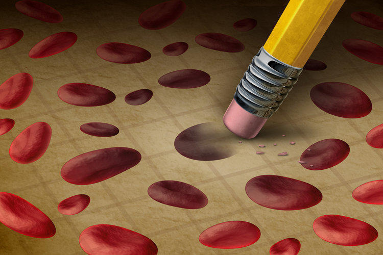
Anemia is a condition of the human body characterized by a low concentration of hemoglobin per unit volume of blood, and, as a rule, correlated with a simultaneous decrease in the number of red blood cells. The state of anemia is secondary and is a symptom of various diseases. Numerous diseases, from diseases of infectious and parasitic etiology to precancerous conditions and the presence of tumors, are accompanied by anemia.
- Anemia in adults
- Causes of anemia in men
- Anemia in women
- Pregnancy Anemia
- Anemia during lactation
- Climacteric Anemia
- Childhood Anemia
- Infant Anemia
- Anemia of Preschool Children
- Anemia in children of primary school age
- Anemia of puberty
- Causes of Anemia
- Classification of Anemia
- Classification by severity
- Classification of varieties by the mechanism of state development
- Color classification
- Morphological classification
- Classification based on assessment of bone marrow’s ability to regenerate
- Iron Deficiency Anemia (IDA)
- Reasons for the development of IDA
- IDA Symptoms
- Diagnosis of IDA
- Treatment of anemia of iron deficiency etiology
- Clinical Nutrition Diet for Iron Deficiency
- Drug Therapy
- Anemia with deficiency of cobalamin or vitamin B12
- Pernicious symptoms
- Diagnosis of cobalamin deficiency
- Treatment methods
- Aplastic anemia: symptoms, causes, diagnosis, treatment
- Causes of the development of aplastic form
- Clinical picture
- Diagnostic Procedures
- Treatment of aplastic form
- Anemia: prevention methods
However, anemia as a condition, causing disturbances in the gas exchange of the body, causes chronic fatigue, increased drowsiness, dizziness, fatigue, increases irritability. In severe cases, anemia can lead to shock states, severe hypotension, coronary, pulmonary insufficiency, hemorrhagic shock. When anemia is detected, treatment is aimed at eliminating the associated symptoms and on treating the underlying disease that caused the state of anemia.
Anemia in adults
Anemia is considered one of the most common pathological conditions among the population of the planet. Among the types of anemia there are several major conditions, classifying them by the causes of anemia:
- iron deficiency anemia;
- hemolytic anemia;
- aplastic anemia;
- sideroblastic type of anemia;
- B12-deficient, resulting from vitamin B12 deficiency;
- post-hemorrhagic anemia;
- sickle cell anemia and other forms.
Approximately one in four people on the planet, according to research by experts, suffers from an iron deficiency anemia due to a decrease in iron concentration. The danger of this condition is in the erased clinical picture of iron deficiency anemia. Symptoms become pronounced when the level of iron and, accordingly, hemoglobin, decreases to a critical point.
The following categories of people are at risk for developing anemia in adults:
- followers of vegetarian food principles;
- people suffering from blood loss due to physiological causes (heavy menstruation in women), diseases (internal bleeding, hemorrhoids, etc.), as well as donors who donate blood and plasma on a regular basis;
- pregnant and lactating women;
- professional athletes;
- patients with chronic or acute forms of certain diseases;
- categories of people who lack nutrition or have a limited diet.
The most common iron deficiency anemia is a consequence of iron deficiency, which, in turn, can be triggered by one of the following factors:
- iron deficiency in food;
- increased need for iron in mind of situational or individual characteristics (developmental pathologies, dysfunctions, diseases, physiological conditions of pregnancy, lactation, professional activity, etc.);
- increased iron loss.
Mild forms of anemia, as a rule, can be cured by correcting the diet, prescribing vitamin-mineral complexes, iron-containing preparations. Medium and severe anemia requires the intervention of a specialist and a course of appropriate therapy.
Causes of anemia in men
The diagnostic criterion for anemia in men is a decrease in hemoglobin concentration in the blood to below 130 g / l. Statistically, anemia in men is less frequently diagnosed than in females due to physiological characteristics: lack of menstruation leading to monthly blood loss, gestation, lactation, often accompanied by deficiencies of essential trace elements.
However, anemia among the male part of the population is also often diagnosed, and, as a rule, is a consequence of the presence of a chronic disease and disorders in the work of various body systems.
Thus, chronic gastric deficiency anemia in men is most often caused by latent gastrointestinal bleeding during intestinal erosion, peptic ulcer, hemorrhoids. In the etiology of anemia in men, there may also be parasitic diseases, benign and malignant neoplasms. A variety of factors that cause anemia require diagnosis of the cause of the condition and appropriate therapy.
Anemia in women
Anemia in women is diagnosed with hemoglobin levels below 120 g / l (or 110 g / l when carrying a child). Physiologically, women are more prone to anemia.
With monthly menstrual bleeding, the female body loses red blood cells. The average volume of monthly blood loss is 40-50 ml of blood, however, with heavy menstruation, the amount of discharge can reach up to 100 ml or more over a period of 5-7 days. A few months of this kind of regular blood loss can lead to anemia.
Another form of hidden anemia that is common among the female population with a high frequency (20% of women) is triggered by a decrease in the concentration of ferritin, a protein that functions as an accumulation of iron in the blood and releases it when the level of hemoglobin decreases.
Pregnancy Anemia

Anemia of pregnant women occur under the influence of various factors. The growing fetus removes from the maternal bloodstream the substances necessary for development, including iron, vitamin B12, folic acid, necessary for the synthesis of hemoglobin. With insufficient intake of vitamins and minerals from food, violations of its processing, chronic diseases (hepatitis, pyelonephritis), pronounced toxicosis of the first trimester, as well as in multiple pregnancies, the expectant mother develops anemia.
The physiological anemia of pregnant women refers to hydremia, blood thinning: in the second half of the gestational period, the volume of the liquid part of the blood increases, which leads to a natural decrease in the concentration of red blood cells and iron transported by them. This condition is normal and is not a sign of pathological anemia, if the hemoglobin level does not fall below 110 g / l or is restored on its own within a short time, and there are no signs of deficiency of vitamins and microelements.
Severe anemia in pregnant women is threatened by miscarriage, premature labor, third trimester toxicosis (gestosis, preeclampsia), complications of the delivery process, and anemia in the newborn.
Symptoms of anemia in pregnant women include the overall clinical picture of anemia (fatigue, drowsiness, irritability, nausea, dizziness, dry skin, brittle hair), as well as distortion of smell and taste (desire to eat chalk, plaster, clay, raw meat, smell substances with a sharp smell among household chemicals, building materials, etc.).
Minor anemia of pregnant and lactating is restored after childbirth and the end of the lactation period. However, with a small gap between repeated births, the recovery process of the body does not have time to complete, which leads to increased signs of anemia, especially pronounced when the interval between births is less than 2 years. The optimal recovery period of the female body is 3-4 years.
Anemia during lactation
According to research by specialists, lactational anemia is most often diagnosed at a fairly pronounced stage of the disease. The development of anemia is associated with blood loss in the process of delivery and lactation on the background of a nursing hypoallergenic diet. By itself, the production of breast milk does not contribute to the development of anemia, however, with the exclusion from the diet of some important food groups, such as legumes (due to the risk of increased gas formation in the child), dairy and meat products (due to allergic reactions in the infant) the likelihood of developing anemia increases significantly.
The reason for the late diagnosis of postpartum anemia is considered to be a shift in the focus of attention from the state of the mother to the child, especially in the youngest mother. The peculiarities of the baby’s health worry her more about her well-being, and the symptom complex of anemia – dizziness, fatigue, drowsiness, decreased concentration, paleness of the skin – is most often perceived as a result of overwork associated with caring for a newborn.
Another reason for the prevalence of iron deficiency anemia of lactating is associated with the wrong opinion about the effect of iron supplements that penetrate into breast milk on the work of the gastrointestinal tract of an infant. This opinion is not confirmed by specialists, and, in the diagnosis of iron deficiency anemia, the medicines and vitamin-mineral complexes prescribed by a specialist are obligatory to receive.
Climacteric Anemia
Anemia during female menopause is a fairly common occurrence. Hormonal restructuring, the effects of menstruation, gestation, childbirth, various dysfunctional conditions and surgical interventions cause chronic anemia, aggravated by climacteric changes in the body.
The provocative role is also played by the restriction in nutrition, unbalanced diets resorted to by women seeking to reduce the rate of weight gain caused by fluctuations in the hormone balance in the pre-menopause and directly during menopause.
By the age of menopause, there is also a decrease in the amount of ferritin in the body, which is an additional factor in the development of anemia.
Fluctuations of well-being, fatigue, irritability, dizziness are often perceived as symptoms of the onset of menopause, which leads to a late diagnosis of anemia.
Childhood Anemia
According to research by the World Health Organization (WHO), 82% of children suffer from anemia of varying severity. Low levels of hemoglobin and iron deficiency of various etiologies lead to impaired mental and physical development of the child. The main causes of anemia in childhood include:
- lack of a full, balanced diet;
- disorders of iron absorption in the gastrointestinal tract;
- dysfunctional regulation of vitamin metabolism;
- parasitic diseases;
- dysbacteriosis, gastritis, gastroduodenitis and other gastrointestinal diseases;
- hormonal imbalances;
- environmental factors: heavy metal poisoning, air pollution, water, food pollution, etc.
The need for iron varies in children depending on age, and upon reaching the puberty period correlates with gender. Therapy of deficient anemia in children with a balanced diet is not always effective, so experts prefer regulation with medications that guarantee the delivery of the required dose of trace elements in the child’s body.
Infant Anemia
A newborn baby is born with a certain amount of iron derived from the mother’s body during fetal development. The combination of the imperfections of one’s own blood formation and rapid physical growth leads to a physiological decrease in the level of hemoglobin in the blood of healthy children who were born in a timely manner, by 4–5 months of life, and by preterm babies – by the age of 3 months.
Artificial and mixed feeding are considered risk factors that increase the likelihood of developing anemia. Hemoglobin deficiency is especially rapidly developing when breast milk and / or artificial mixtures are replaced by cow, goat milk, cereals and other products in the period up to 9-12 months.
Symptoms of anemia in children under one year include:
- pallor of the skin, since the skin is still very thin, there is increased “transparency”, “cyanosis” of the skin;
- worry, causeless crying;
- sleep disorders;
- loss of appetite;
- hair loss outside the physiological framework of changing hair;
- frequent regurgitation;
- low weight gain;
- lagging first in physical, then in psycho-emotional development, loss of interest, inexpressiveness of the animation complex, etc.
The peculiarity of children of this age is the ability for high (up to 70%) absorption of iron from food, therefore, not all cases of anemia, pediatricians see the need for prescribing drugs, limited to correcting the infant’s diet, transferring to full breastfeeding, and matching the needs substitute. In case of severe anemia, iron preparations are prescribed in the age dosage, for example, Ferrum Lek or Maltofer in the form of syrup drops.
When diagnosing a severe degree of anemia, the reasons may not be in the diet, but in diseases, pathologies and dysfunctions of the child’s body. Anemia can also be caused by hereditary diseases, some hereditary developmental disorders and diseases are characterized by a decrease in iron concentration, spectrocytopenia, insufficiency of the hematopoietic system, etc. With persistent low hemoglobin levels, a mandatory examination of children and correction of the primary disease are necessary.
Anemia of Preschool Children
Conducted in 2010, a large-scale study revealed a high frequency of the presence of iron deficiency anemia in preschool children: every second child suffers from a lack of hemoglobin due to low levels of iron. There are various factors in the etiology of this phenomenon, but the most common is the consequences of uncorrected anemia in the first year of life.
The second factor that provokes anemia in preschoolers is often combined with the first. Lack of a balanced diet, lack of protein (meat products) and vitamins (vegetables) is often explained by the child’s reluctance to eat meat and vegetables, preferring semi-finished products and sweets. It is solely a question of educating parents and attention to a healthy diet without providing alternative products from an early age, which also requires the transfer of family members to a rationally composed diet.
In the case when the diet meets age standards, and the child shows signs of anemia (pallor, dry skin, fast fatigue, reduced appetite, increased fragility of the nail plates, etc.), a specialist should be examined. Despite the fact that in 9 out of 10 preschool children with diagnosed anemia, it is due to iron deficiency, in 10% of anemia, the cause is in diseases and pathologies (celiac disease, leukemia, etc.).
Anemia in children of primary school age
Norms of hemoglobin in the blood of children 7-11 years old – 130 g / l. Manifestations of anemia in this age period increase gradually. Signs of developing anemia include, in addition to the symptoms of anemia in preschool children, a decrease in concentration, frequent acute respiratory viral and bacterial diseases, fatigue, which may affect the results of educational activity.
An important factor in the development of anemia in children attending general education institutions is the lack of control over diet. In this age period, a sufficient level of iron absorption from food entering the body is maintained (up to 10%, decreasing to 3% by an adult’s age); therefore, a well-organized meal with rich vitamins and microelements at its core serves as prevention and correction of iron deficiency anemia.
Hypodynamia, limited outdoor exposure, preference for games in the house, especially with tablets, smartphones, etc., which dictate a long stay in a static position, also provoke anemia.
Anemia of puberty
Adolescence is dangerous for the development of anemia, especially in girls with the onset of menstruation, characterized by a periodic decrease in hemoglobin with blood loss. The second factor provoking the onset of anemia in adolescent girls is associated with concentrating on one’s own appearance, striving to adhere to various diets and reducing the daily diet, eliminating the necessary health products.
Rapid growth, intense sports, poor nutrition and anemia of the previous period also affect adolescents of both sexes. Symptoms of anemia in adolescence include a blue tinge to the sclera of the eyes, a change in the shape of the nails (the cup-shaped shape of the nail plate), dysfunction of the digestive system, taste disorders, smell.
Pronounced forms of the disease in adolescence require medication therapy. A change in the blood formula is noted, as a rule, not earlier than 10-12 days after the start of the course of treatment, signs of clinical recovery, subject to compliance with the prescription, are observed after 6-8 weeks.
Causes of Anemia
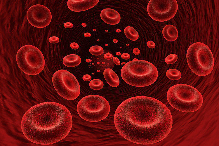
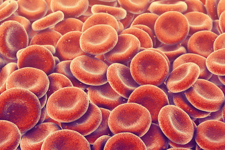
Anemia is characterized by a decrease in the concentration of hemoglobin and red blood cells per unit of blood. The main purpose of red blood cells is to participate in gas exchange, transport of oxygen and carbon dioxide, as well as nutrients and metabolic products to cells and tissues for further processing.
The erythrocyte is filled with hemoglobin, a protein that gives the erythrocyte and blood red color. The composition of hemoglobin includes iron, and therefore its lack in the body causes a high frequency of iron-deficient varieties of anemia among all the varieties of this condition.
There are three main factors for the development of anemia:
- acute or chronic blood loss;
- hemolysis, red blood cell destruction;
- reduced bone marrow production of red blood cells.
For a variety of factors and causes, the following types of anemia are distinguished:
- food associated with a dietary deficiency or a general lack of food;
- physical (trauma, surgery, delivery, frostbite, burns, etc.);
- genetic etiology;
- infectious, secondary anemia in such diseases as viral hepatitis, cirrhosis, tuberculosis of the liver, glomerulonephritis, gastrointestinal tract diseases (peptic ulcer, gastritis, Crohn’s disease), rheumatoid arthritis, systemic lupus, benign and malignant neoplasms different localization;
- infectious (with viral, bacterial, parasitic and protozoal diseases);
- poisoning with poisonous substances and medicines, including during a long, especially uncontrolled course of therapy (antibiotic therapy, taking cytotoxic drugs, nonsteroidal anti-inflammatory drugs, antithyroid, antiepileptic drugs);
- exposure to radioactive waves.
Classification of Anemia
The classification of an anemic condition is based on various signs describing the etiology, mechanisms of disease development, the stage of anemia, diagnostic indicators.
Classification by severity
The severity of anemia is based on blood test results and depends on the age, gender and physiological period.
Normally, in a healthy adult male, hemoglobin values are 130-160 g / l of blood, in a woman – from 120 to 140 g / l, in the gestation period from 110 to 130 g / l.
A mild degree is diagnosed when the level of hemoglobin concentration is up to 90 g / l in both sexes, with an average figure corresponding to the range from 70 to 90 g / l, severe anemia is characterized by a decrease in hemoglobin level below the 70 g / l limit.
Classification of varieties by the mechanism of state development
In the pathogenesis of anemia, there are three factors that can act separately or together:
- acute or chronic blood loss;
- disorders of the hematopoietic system, the production of red blood cells by the bone marrow (iron deficiency, renal, aplastic anemia, deficient anemia with a deficiency of vitamin B12 and / or folic acid);
- increased destruction of red blood cells before the end of the term of operation (120 days) due to genetic factors, autoimmune diseases.
Color classification
The color indicator serves as an indicator of the erythrocyte saturation with hemoglobin and is calculated using a special formula in the blood analysis process.
Hypochromic form with a weak red blood cell color is diagnosed with a color index below 0.80.
The normochromic form, with a color index within the normal range, is determined by the range 0.80-1.05.
The hyperchromic form, with excessive hemoglobin saturation, corresponds to a color index higher than 1.05.
Morphological classification
The size of red blood cells is an important indicator in diagnosing the causes of anemia. Different sizes of red blood cells may indicate the etiology and pathogenesis of the condition. Normally, red blood cells are produced with a diameter of 7 to 8.2 micrometers. The following species are distinguished on the basis of determining the size of the prevalent red blood cell count in the blood:
- microcytic, red blood cell diameter less than 7 microns, indicates a high probability of iron deficiency;
- normocytic variety, the size of red blood cells from 7 to 8.2 microns. Normocytosis is a sign of post-gemological form;
- macrocytic, with a red blood cell size of more than 8.2 and less than 11 microns, as a rule, indicates a deficiency of vitamin B12 (pernicious form) or folic acid;
- megalocytosis, megalocytic (megaloblastic) form in which the diameter of erythrocytes is more than 11 μm, corresponds to the severe stages of some forms, disturbances in the formation of red blood cells, etc.
Classification based on assessment of bone marrow’s ability to regenerate
The degree of erythropoiesis, the ability of the red bone marrow to form red blood cells, is assessed by the quantitative indicator of reticulocytes, progenitor cells or “immature” erythrocytes, which is considered the main criterion for assessing the ability of bone marrow tissue to regenerate and is an important factor for predicting the patient’s condition and choice of therapy methods. The normal concentration of reticulocytes is an indicator of 0.5-1.2% of the total number of erythrocytes per unit of blood.
Depending on the level of reticulocytes, the following forms are identified:
- regenerative, indicating normal bone marrow’s ability to recover. The level of reticulocytes 0.5-1.2%;
- Hyporegenerative, with a concentration of immature red blood cells below 0.5%, which indicates a reduced ability of the bone marrow to restore itself;
- hyperregenerator, reticulocyte rate more than 2%;
- Aplastic anemia is diagnosed when the concentration of immature red blood cells is less than 0.2% among the mass of all red blood cells and is a sign of a sharp suppression of the ability to regenerate.
Iron Deficiency Anemia (IDA)
The iron deficiency makes up to 90% of all varieties of anemic conditions. According to the World Health Organization, one out of six men and one out of every three women in the world suffer from this form.
Hemoglobin is a complex protein compound that has iron in ts composition and is capable of reversibly binding to oxygen molecules, which is the basis of the process of transporting oxygen from the lungs to the body’s tissues.
Iron deficiency is hypochromic anemia, with signs of microcytosis, red blood cells with a diameter less than normal in the blood formula, which is associated with iron deficiency, a basic element for the formation of hemoglobin that fills the red blood cavity.
Iron is a vital trace element involved in many metabolic processes, nutrient exchange, and gas exchange of the body. During the day, an adult consumes 20-25 mg of iron, while the total stock of this element in the body is about 4 g.
Reasons for the development of IDA
The reasons for the development of this form of condition include factors of different etiologies.
Iron damage:
- unbalanced diet, strict vegetarianism without compensating for iron-containing foods, starvation, diets, taking medicines, drugs and other substances that suppress hunger, anorexia due to physical or psycho-emotional diseases;
- Socio-economic causes of malnutrition, lack of food.
Disorders of the absorption process, the absorption of iron:
- diseases of the gastrointestinal tract (gastritis, colitis, gastric ulcer, resection of the organ).
Imbalance of consumption and iron supply due to increased body needs:
- pregnancy, lactation;
- age of pubertal physical growth jumps;
- chronic diseases provoking hypoxia (bronchitis, obstructive pulmonary disease, heart defects and other diseases of the cardiovascular system and respiratory organs);
- diseases involving purulent-necrotic processes: sepsis, tissue abscesses, bronchiectasis, etc.
Iron loss by the body, acute or chronic post-hemorrhagic:
- for pulmonary hemorrhage (tuberculosis, tumor formation in the lungs);
- for gastrointestinal bleeding accompanying gastric ulcer, duodenal ulcer, gastric and intestinal cancers, marked erosion of the gastrointestinal mucosa, varicose veins of the esophagus, rectum, hemorrhoids, intestinal worm ulcer, nonspecific ulcerative colitis and others;
- for uterine bleeding (heavy menstruation, cancer of the uterus, cervix, fibroids, placental abruption in the gestational period or in childbirth, ectopic pregnancy during exile, birth injuries of the uterus and cervix);
- bleeding with localization in the kidney (tumor formation in the kidney, tuberculous changes in the kidney);
- bleeding, including internal and hidden, due to injuries, blood loss from burns, frostbite, planned and emergency surgery, etc.
IDA Symptoms
The clinical picture of the iron deficiency is made up of an anemic and sideropenic syndrome, caused primarily by insufficient gas exchange in the body’s tissues.
Symptoms of anemic syndrome include:
- general malaise, chronic fatigue;
- weakness, inability to endure prolonged physical and mental stress;
- attention deficit disorder, difficulty concentrating, rigidity;
- irritability;
- headaches;
- dizziness, sometimes fainting;
- drowsiness and sleep disorders;
- dyspnea, rapid heart rate, both during physical and / or psycho-emotional stress, and at rest;
- black stools (for bleeding in the gastrointestinal tract).
Sideropenic syndrome is characterized by the following manifestations:
- perversion of taste preferences, craving for eating chalk, clay, raw meat, etc.;
- olfactory distortion, the desire to smell paint, household chemicals, substances with a pungent odor (acetone, gasoline, washing powder, etc.);
- brittle, dry hair, no shine;
- white spots on the nail plates of the hands;
- dry skin, peeling;
- paleness of the skin, sometimes blue sclera;
- the presence of cheilitis (cracks, “zade”) in the corners of the lips.
In severe stages of IDA, neurological symptoms are observed: goose bumps, numbness of the limbs, difficulty in swallowing, weakening of bladder control, etc.
Diagnosis of IDA
The diagnosis of iron deficiency anemia is based on external examination, assessment of the results of laboratory blood tests and instrumental examination of the patient.
During external medical examination and collection of anamnesis, attention is paid to the condition of the skin, mucous surfaces of the mouth, the corners of the lips, and also the size of the spleen on palpation is assessed.
Complete blood count in the classical clinical picture of IDA shows a decrease in the concentration of red blood cells and hemoglobin relative to age and sex norms, the presence of red blood cells of various sizes (poikilocytosis), reveals microcytosis, the presence, in severe forms, the predominance of red blood cells with a diameter of less than 7.2 microns, hypochromic , poorly expressed color of erythrocytes, low color index.
The results of biochemical blood tests for IDA have the following indicators:
- low concentration of ferritin, a protein that acts as a depot of iron in the body;
- low serum iron;
- increased serum iron-binding ability.
Diagnosis of IDA is not limited to detecting iron deficiency. For effective correction of the condition after anamnesis is collected, a specialist, if necessary, assigns instrumental studies to clarify the pathogenesis of the disease. In this case, instrumental studies include:
- fibrogastroduodenoscopy, examination of the state of the mucous membrane of the esophagus, the walls of the stomach, duodenum;
- ultrasound examination of the liver, kidneys, female reproductive organs;
- colonoscopy, examination of the walls of the large intestine;
- computed tomography methods;
- X-ray examination of the lungs.
Treatment of anemia of iron deficiency etiology
Depending on the stage and pathogenesis of IDA, therapy is chosen through dietary correction, medication, surgery to eliminate the causes of blood loss or a combination of methods.
Clinical Nutrition Diet for Iron Deficiency
Iron entering the body with food is divided into heme, animal, and non-heme iron of plant origin. Heme variety is absorbed much better and its lack of nutrition, for example, in vegetarians, leads to the development of IDA.
The products recommended for correcting iron deficiency include the following:
- heme group in order of decreasing amounts of iron: beef liver, beef tongue, rabbit, turkey, goose meat, beef, some varieties of fish;
- non-heme group: dried mushrooms, fresh peas, buckwheat, oats and oats, fresh mushrooms, apricots, pears, apples, plums, cherries, beets, etc.
Despite the apparent high in the study of the composition of iron in vegetables, fruits, products of plant origin, the absorption of iron from them is insignificant, 1-3% of the total, especially comparing with products of animal origin. Thus, when eating beef, the body is able to absorb up to 12% of the necessary element contained in meat.
When correcting IDA using a diet, one should increase the content of foods rich in vitamin C and protein (meat) in the diet and reduce the consumption of eggs, table salt, caffeinated beverages and foods rich in calcium due to the effect on the digestion of dietary iron.
Drug Therapy
In moderate and severe forms, a therapeutic diet is combined with prescribing iron-supplying drugs in an easily digestible form. Medicines differ in the type of compound, dosage, and release form: tablets, dragees, syrups, drops, capsules, solutions for injections.
Preparations for oral administration are taken one hour before meals or two hours after due to the absorption of iron, while caffeine-containing beverages (tea, coffee) are not recommended as a fluid that facilitates ingestion, since this impairs the absorption of the element. The interval between doses of drugs should be at least 4 hours. Independent administration of medications can cause both side effects from an incorrectly chosen form or dosage, and iron poisoning.
The dosage of drugs and the form of release is determined by the specialist, focusing on the age, stage of the disease, causes of the condition, general clinical picture and individual characteristics of the patient. Doses can be adjusted during the course of treatment according to the results of intermediate or control blood tests and / or the patient’s well-being.
Iron preparations in the course of treatment take from 3-4 weeks to several months with periodic monitoring of hemoglobin levels.
Among the drugs supplying iron, taken orally, emit drugs with two – and trivalent form of iron. At the moment, according to research, bivalent iron is considered to be the preferred form for oral administration due to a higher ability to be absorbed in the body and a gentle effect on the stomach.
For children, they produce iron-containing agents in the form of drops and syrups, which is caused by both the age-related features of taking the drugs and the shorter course of therapy than in adults, due to the increased digestibility of iron coming from food. If you can take capsules, dragees and tablets, as well as during long courses, you should prefer solid forms of medicines containing iron, since the liquid with prolonged use can have a negative effect on the tooth enamel and cause darkening.
The most popular pill forms include the following medicines: Ferroplex, Sorbifer, Aktiferrin, Totem (ferrous iron form) and Maltofer, Ferrostat, Ferrum Lek with trivalent iron.
Oral forms are combined with the intake of vitamin C (ascorbic acid) in the dosage prescribed by the doctor for better absorption.
Intramuscular and intravenous injections of iron supplements are prescribed in limited situations, such as:
- severe anemia;
- the ineffectiveness of the course of taking oral forms of drugs;
- the presence of specific diseases of the gastrointestinal tract, in which oral administration can worsen the patient’s condition (in acute gastritis, gastric ulcer, duodenal ulcer, ulcerative colitis, Crohn’s disease, etc.);
- with individual intolerance to oral forms of iron-containing drugs;
- in situations of urgent need to saturate the body with iron, for example, with significant blood loss due to injury or before surgery.
The introduction of iron preparations intravenously and intramuscularly can lead to an intolerance reaction, which is why a similar course of therapy is carried out exclusively under the supervision of a specialist in a hospital or clinical setting. The adverse effects of intramuscular injection of iron-containing fluids include the deposition of hemosiderin subcutaneously at the injection site. Dark spots on the skin at the injection sites can last from one and a half to 5 years.
Iron deficiency anemia responds well to drug therapy, subject to the prescribed dose and duration of treatment. However, if the primary etiology of the condition includes primary serious diseases and disorders, the therapy will be symptomatic and have a short-term effect.
To eliminate such causes as internal bleeding, in hemorrhagic form, iron deficiency anemia is treated by surgical methods. Surgical intervention allows to eliminate the main factor of acute or chronic bleeding, to stop blood loss. For internal bleeding of the gastrointestinal tract, fibrogastroduodenoscopic methods or colonoscopy are used to identify the area of bleeding and measures to stop it, for example, cutting off a polyp, coagulating the ulcer.
For internal bleeding of the peritoneum and reproductive organs in women, laparoscopic intervention is used.
Emergency treatment methods include the transfusion of donor red blood cells to quickly restore the level of red blood cells and hemoglobin per unit of blood.
Prevention of iron deficiency is considered a balanced diet and timely diagnostic and therapeutic measures to preserve health.
Anemia with deficiency of cobalamin or vitamin B12
Deficits are not limited to iron deficiency anemia. Pernicious anemia is a condition that occurs on the background of a suction violation vitamin B12, its inadequate intake, increased consumption, deviations in the synthesis of protective protein or pathologies of the liver, preventing the accumulation and storage of cobalamin. In ptogenesis of this form, a frequent combination with folic acid deficiency is also noted.
Among the causes of this deficient form are the following:
- Vitamin B12 deficiency in food. Normally, the liver contains reserves of cobalamin, capable of meeting the needs of the body for 2-4 years. For the food factor, vitamin B12 deficiency must be pronounced and prolonged (fasting, a monotonous diet);
- violations of the synthesis of internal factor Castle or gastromucoprotein, a protein that protects cobalamin from the negative effects of intestinal flora and participating in the absorption of vitamin by the intestinal walls. This deviation can be observed in diseases of the gastrointestinal tract (atrophic gastritis, gastrectomy, tumors of the stomach and intestines);
- intestinal dysfunction due to pronounced dysbiosis, parasitosis, worm infestations, intestinal infectious diseases;
- increased body’s need for cobalamin: gestation period, especially during multiple pregnancies, the stage of rapid growth (infancy, puberty), excessive exercise without correction of nutrition to the needs of the body;
- a decrease in the liver’s depositing function due to diseases affecting the structure of its tissues, for example, cirrhosis.
Pernicious symptoms
The clinical picture of vitamin B12 deficiency and folic acid include anemic, gastrointestinal, and neuralgic syndromes.
Particularly of the anemic symptom complex with this type of deficiency includes such specific symptoms as yellowness of the skin and sclera and an increase in blood pressure. Other manifestations are characteristic including for IDA: weakness, fatigue, dizziness, shortness of breath, rapid heartbeat (situationally), tachycardia, etc.
The symptoms associated with the functioning of the gastrointestinal tract, include the following symptoms of atrophy of the mucous membranes of the gastrointestinal tract and oral cavity:
- red, “glossy” language, often with complaints of a burning sensation of its surface;
- the phenomena of aphthous stomatitis, ulceration of the oral mucosa;
- appetite disturbances: decrease until complete absence;
- feeling of heaviness in the stomach after eating;
- weight loss in the patient in the immediate history;
- violations, difficulties in the process of defecation, constipation, pain in the rectum;
- hepatomegaly, liver enlargement in size.
Neuralgic syndrome with vitamin B12 deficiency consists of the following manifestations:
- feeling of weakness in the lower limbs with severe physical exertion;
- numbness, tingling, “goosebumps” on the surface of the arms and legs;
- reduced peripheral sensitivity;
- atrophy of the muscular tissue of the legs;
- Convulsive manifestations, muscle cramps, etc.
Diagnosis of cobalamin deficiency
Diagnostic measures include general medical examination of the patient, history taking, laboratory blood tests and, if necessary, instrumental examination methods.
With a general blood test, the following changes are noted:
- Erythrocyte and hemoglobin levels decreased relative to the age norm limits;
- hyperchromia, increased red blood cell color index;
- macrocytosis of erythrocytes, exceeding their size in diameter of more than 8.0 microns;
- poikilocytosis, the presence of red blood cells of various sizes;
- leukopenia, insufficient concentration of white blood cells;
- lymphocytosis, exceeding the limits of normal levels of lymphocytes in the blood;
- thrombocytopenia, insufficient platelet count per unit of blood.
Blood tests using biochemistry reveal hyperbilirubinemia and vitamin B12 deficiency.
To diagnose the presence and severity of atrophy of the mucous membranes of the stomach and intestines, as well as to identify possible primary diseases using instrumental methods of examination of patients:
- fibrogastroduodenoscopy;
- analysis of biopsy material;
- colonoscopy;
- irrigoscopy;
- liver ultrasound.
Treatment methods
In most cases, B12-deficient anemia requires hospitalization or inpatient treatment. For therapy, first of all they prescribe a diet with foods saturated with cobalamin and folic acid (liver, beef, mackerel, sardines, cod, cheese, etc.), secondly they use medicamental support.
In the presence of neurological symptoms, cyanocobalamin injections are administered intramuscularly in an overdose: 1000 mcg daily until the neurological signs of deficiency disappear. In the future, the dosage is reduced, however, with a diagnosis of secondary etiology, the medication is most often prescribed on a lifelong basis.
After discharge from the hospital, the patient must undergo regular routine check-ups with a therapist, hematologist and gastrologist.
Aplastic anemia: symptoms, causes, diagnosis, treatment
Aplastic anemia can be both congenital and acquired disease, developing under the influence of internal and external factors. The condition itself is caused by bone marrow hypoplasia, a decrease in the ability to produce blood cells (red blood cells, white blood cells, platelets, lymphocytes).
Causes of the development of aplastic form
In aplastic, hypoplastic forms of anemia, the causes of this condition may be as follows:
- stem cell defect;
- suppression of the process of hemopoiesis (blood formation);
- insufficiency of hematopoietic stimulation factors;
- immune, autoimmune reactions;
- iron deficiency, vitamin B12 or their exclusion from the process of hemopoiesis due to impaired function of blood-forming tissues and organs.
The development of disorders that provoke the aplastic or hypoplastic form includes the following factors:
- hereditary diseases and genetic pathologies;
- taking certain medications from antibiotic groups, cytostatics, nonsteroidal anti-inflammatory drugs;
- chemical poisoning (benzenes, arsenic, etc.);
- infectious diseases of viral etiology (parvovirus, human immunodeficiency virus);
- autoimmune disorders (systemic lupus erythematosus, rheumatoid arthritis);
- severe deficiencies of cobalamin and folic acid in the diet.
Despite the extensive list of causes of the disease, in 50% of cases the pathogenesis of the aplastic form remains undetected.
Clinical picture
The severity of pancytopenia, reducing the number of basic types of blood cells, determines the severity of symptoms. The clinical signs of the aplastic form include the following signs:
- tachycardia, rapid heartbeat;
- pale skin, mucous;
- headaches;
- increased fatigue, sleepiness;
- shortness of breath;
- lower limb edema;
- bleeding gums;
- petechial rash in the form of small red spots on the skin, a tendency to bruise easily;
- frequent acute infections, chronic diseases as a result of reduced general immunity and leukocyte insufficiency;
- erosion, ulcers on the inner surface of the oral cavity;
- yellowness of the skin, sclera of the eyes as a sign of the onset of liver damage.
Diagnostic Procedures
To establish the diagnosis using laboratory methods for the study of various biological fluids and tissues and instrumental examination.
In the general analysis of the blood, a reduced number of erythrocytes, hemoglobin, reticulocytes, leukocytes, and platelets is noted when the norm is in a color index and hemoglobin content in erythrocytes. The results of the biochemical studies indicate an increase in serum iron, bilirubin, lactate dehydrogenase, transferrin saturation with iron by 100% of the possible.
To clarify the diagnosis, histological examination of the material removed from the bone marrow during puncture is performed. As a rule, according to the results of the study, underdevelopment of all shoots is noted and bone marrow is replaced with fat.
Treatment of aplastic form
Anemia of this variety is not treatable with diet correction. First of all, patients with aplastic anemia are prescribed selective or combined medication in the following groups:
- immunosuppressants;
- glucocorticosteroids;
- anti-lymphocyte and anti-platelet immunoglobulins;
- antrimetabolic drugs;
- erythrocyte stem cell stimulants.
With the ineffectiveness of drug therapy, non-drug treatment methods are prescribed:
- bone marrow transplantation;
- red blood cell platelet transfusion;
- plasmapheresis.
Aplastic anemia is accompanied by a decrease in general immunity due to leukocyte deficiency, therefore, in addition to general therapy, aseptic environment, antiseptic surface treatment, and no contact with carriers of infectious diseases are recommended.
In case of insufficiency of the listed methods of treatment, the patient is prescribed splenectomy surgery, removal of the spleen. Since it is in this organ that the red blood cells break down, its removal can improve the patient’s general condition and slow the progression of the disease.
Anemia: prevention methods
The most common form of the disease – iron deficiency anemia – is to be prevented by using a balanced diet with an increase in the amount of iron-containing foods during critical periods. An important factor is the presence in food vitamin C, cobalamin (vitamin B12), folic acid.
If you are at risk of developing this form of anemia (vegetarianism, age periods of growth, pregnancy, lactation, prematurity in infants, heavy menstrual bleeding, chronic and acute diseases), regular medical examinations, blood tests for quantitative and qualitative hemoglobin, red blood cells and additional taking drugs in accordance with the appointment of specialists.