Among all cancers to skin cancer, perhaps, many have the most frivolous attitude. Not all people represent how dangerous he can be. But very often skin cancer can be recognized in the early stages, when it is very easy to cure. Therefore, seeing unusual formations on your skin, you should immediately go to the doctor. But in what cases is there a cause for concern, and in what is not?
- Risk Factors
- What does skin cancer look like
- First signs of skin cancer on the body (initial stage)
- Basal cell carcinoma of the skin
- Nodular (nodular, solid) basalioma
- Superficial basalioma
- Scleroderma-like (flat, morphe-like, sclerosing) basalioma
- Cystic Basalioma
- Fibroepithelial basalioma (Pincus fibroepithelioma)
- Squamous cell cancer
- Tumors of skin appendages
- Adenocarcinoma
- Verrucous Carcinoma
- Pre-cancerous conditions of the skin
- Bowen’s Disease
- Cutaneous Horn
- Keratoacanthoma
- Actinic keratosis
- Dysplastic Nevus
- Diagnosis of the disease
- Treatment
There are many types of malignant skin lesions, and all of them are significantly different both in nature and in the severity of the disease. Some types of skin cancer are very rare or in certain categories of the population, while others may be sick for people of different gender and age.
Unlike some other types of oncological diseases that are capable of implicitly developing in the early stages without special symptoms, skin cancer in the early stages is usually easy to notice. After all, the surface of the skin is almost always available for visual review. This means that a person is able to pay attention to the reborn tissue.
Risk Factors
Why does cancer develop, particularly on the skin? Medicine has no definite answer to this question. Undoubtedly, not only one unfavorable factor plays its role here, but their combination at once. Scientists believe that the following circumstances contribute most to the development of tumors:
- smoking;
- unhealthy lifestyle;
- lack of personal hygiene;
- unhealthy diet, consuming large amounts of potentially carcinogenic foods and not enough vitamins and fiber in the diet;
- injuries and injuries to the surface of the skin;
- hereditary factors;
- racial features;
- long exposure to solar radiation;
- frequent use of tanning beds;
- exposure to ionizing radiation;
- prolonged contact with potentially carcinogenic substances (soot, fuel oil, benzene, coal tar, oil, etc.);
- outdoor work;
- advanced age (over 50);
- long-term use of corticosteroids and immunosuppressants;
- low immunity;
- high incidence of other types of skin pathologies;
- prolonged exposure to high temperature;
- precancerous conditions of the skin (optional and obligate)
- systemic lupus erythematosus;
- AIDS;
- chemotherapy and radiation therapy for other cancers;
- hormonal changes (including during pregnancy);
For different types of skin cancer, the proportion of individual factors may not be the same. For example, some species can manifest themselves almost exclusively in old age. However, one way or another, almost all types of skin cancer are observed mainly in adulthood. Cases of the disease of children are relatively rare. The frequency of other types of malignant tumors varies greatly depending on racial and sexual factors.
What does skin cancer look like
Different types of skin cancer may look different. However, no matter what kind of skin cancer a person has, the symptoms of the disease may be similar:
- burning and itching,
- soreness
- bleeding,
- red border around the tumor.
Phenomena such as darkening of the previously light skin area, long ulceration of the surface, enlargement and soreness of the lymph nodes near the site of the neoplasm, compaction of the skin with its elevation above the surface should also be alarming. The soreness of skin formations may indicate germination of the tumor in the deep, subcutaneous tissue layers or the accession of secondary inflammatory processes.
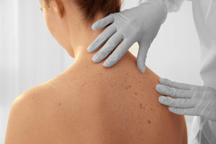
What should I do if there are suspicious signs? First of all, in no case should you postpone a visit to the doctor. After all, the sooner treatment is started, the greater the chance of a happy outcome.
First signs of skin cancer on the body (initial stage)
Malignant neoplasms of the skin are diverse. Their main groups:
- non-melanoma tumors – basal cell and squamous cell carcinomas – develop from epithelial skin cells;
- melanoma;
- tumors of the skin appendages;
- other neoplasms.
Let us describe the first signs of skin cancer of the most common types of malignant tumors.
Basal cell carcinoma of the skin
Basal cell carcinoma of the skin (synonyms – basal cell carcinoma, basal cell carcinoma, Krompeher basal cell carcinoma) develops from the cells of the basal layer of the skin epithelium.
This type of skin cancer is most common (in about 75% of cases). However, in most cases only older people are sick (over 60 years old). Basalioma has the slowest development and most favorable prognosis among all types of skin cancer. Basalioma is located, usually on the skin of the face, most often on the following surfaces:
- nose bridge,
- brow area,
- nose wings,
- temple,
- upper lip,
- nasolabial fold,
Basalioma can also occur on the ears and neck. Growing to large sizes, it can grow through the skin and underlying tissues and cause damage to them. Due to the fact that basal cell carcinoma grows slowly, patients do not immediately consult her to the doctor.
Basalioma usually occurs suddenly, without any precursors to precancerous tumors, unlike squamous cell carcinoma of the skin. The diameter of the tumor in the beginning stage is usually 2 cm, it is easily injured and bleeds.
Unlike other types of skin tumors, basalioma rarely metastasizes. In principle, this type of tumor is something between benign and malignant tumors. However, according to histological features, it still refers to malignant tumors. The prognosis for this category of skin tumors is favorable.
Dangerous with this type of skin cancer are cases where the localization of the pathology occurs around the eyes, in the folds above the lip, around the ear canal, in the posterior sulcus of the auricle. In these places, the tumor grows deep, damaging the bone tissue, muscles, brain.
However, with early detection, timely treatment and removal of the tumor, the patient can get rid of this disease without consequences.
There are about 20 types of basal cell carcinoma cancer cells. The most common clinical forms are:
- node;
- superficial;
- scleroderm-like;
- cystic;
- fibroepithelial.
The symptoms and signs of each type of basal cancer differ. And most often in one patient forms are combined. Diagnosis requires a clinical examination by a specialist doctor.
Nodular (nodular, solid) basalioma
Appears on the scalp, neck. This initial skin cancer is characterized by the appearance of dense small nodules (2-5 mm), which gradually merge with each other. The tumor grows slowly, then disintegrates, forming a deep ulcer with valiform edges, covered with purulent necrotic crusts.
Pigment tumor cells can be translucent, and can vary from slightly brown to black.
Superficial basalioma
Usually located on the torso, arms and legs. Looks like a rounded pink spot. At the initial stage, it peels off, and when developing, papilloma growths and ulcerations appear on its surface.
This skin cancer is not aggressive and most favorable according to the forecast: the defeat of the skin develops very slowly, for decades.
Scleroderma-like (flat, morphe-like, sclerosing) basalioma
Rare, but rather aggressive skin cancer. The tumor is deep in the layers of the skin and by volume it is much larger than the external signs on the surface. There are many cases of disease recurrence.
Most symptoms of this type of skin cancer are found on the head and neck. At the initial stage, a pale pink plaque with raised edges and mother-of-pearl shade appears on the skin. In the later stages of development, the lesion has the form of a depressed scar or patch.
Cystic Basalioma
The form is called cystic, because it looks like a skin cancer of this type of translucent nodule (like a cyst). Detected by chance if a biopsy is performed.
Fibroepithelial basalioma (Pincus fibroepithelioma)
Signs of skin cancer of this type are usually found on the lower back. Looks like a fibrous polyp on a flat or hemispherical leg. This is a rare tumor formation. It has a favorable outlook.
Squamous cell cancer
Squamous cell carcinoma of the skin (synonyms – squamous cell carcinoma, squamous epithelioma, epidermoid carcinoma, spinocellular carcinoma) develops from skin keratinocytes.
This type of skin cancer is the third most common cancer after basal cell carcinoma and melanoma. They can suffer people of any age, both adults and elderly, both men and women.
Externally, the tumor in squamous cancer resembles a small sore, sometimes bleeding. Very often, the tumor is confused with the manifestation of some inflammatory skin diseases, dermatitis, burns. However, unlike these formations, the tumor does not shrink and grows.
This formation can be found in various parts of the body, but is most often found at the junction of various skin surfaces – the corners of the eyes, mouth, lips, mucous membranes, genitals (Keir’s disease), etc. Over time, the tumor may form metastases. However, they are most often observed on the skin near the primary tumor on the skin or in the lymph nodes. For tumors located on the face, damage to the lymphatic system is most common. Signs of lymph nodes are their increase in size, their mobility and soreness. In the future, their decay is possible with simultaneous ulceration of the nearest skin. Metastases affect distant organs only in advanced cases of the disease.
A tumor is usually distinguished by slow development, which increases the chances that it can be promptly recognized and cured. However, at a late stage of development of the disease, the percentage of patient survival is small.
Squamous cell carcinoma in the initial stage is a formation in the form of a red seal, an ulcer or bumps with a diameter of about 2 cm. This formation can easily be injured. The trigger for its development can serve as various factors, first of all, the intense irradiation of the skin by the sun’s rays. On the site of the appearance of education can be as a healthy area of the skin, and scars from burns, chronic ulcers, inflammation.
Squamous cell carcinoma is easily treatable in its early stages. For this purpose, using surgical methods, radiation therapy, chemotherapy. However, chemotherapy in most cases is auxiliary.
As a variety of squamous cell carcinoma of the skin, you can specify a highly differentiated cancer. The predecessor of this disease is such pathological formations as actinic keratosis and Bowen’s disease.
With highly differentiated skin cancer, the tumor grows long. It has a high density, horny growths and crusts on the surface. However, this skin tumor is similar to warts, solar keratosis, which can make it difficult to diagnose the disease.
Low-grade squamous cell carcinoma, unlike well-differentiated, has a high growth rate and an aggressive course. This is a soft formation, having the appearance of a bump or ulcer. May bleed or ache.
Five-year survival with timely removal of cancer cells is more than 50%, but when metastasis forms, it decreases to 30%.
Melanoma
This tumor develops on the basis of the pigment cells of the skin – melanocytes. This type of cancer is relatively rare compared with basal cell (15% of all skin cancer cases). However, he still takes second place after it, and if you take all cancers, more than 1% of them fall on melanoma. Most often, women suffer from the disease, although the proportion of sick men is quite large. The likelihood of this type of skin cancer in people over the age of 50 also increases dramatically.
Despite the relative low probability of developing this type of cancer, it is among the most aggressive types of skin cancer, and cancer in general. For reasons that are not completely understood by science, the body’s immune system reacts very poorly to melanoma, allowing tumors to develop quite quickly — within weeks, and sometimes several days, the tumor goes from cancer in the initial stage to the life-threatening stage. Also, the tumor is characterized by rapid metastasis in the early stages, and metastases can penetrate not only the areas of the skin adjacent to the tumor, but also to the lymph nodes, as well as organs distant from the tumor.
The prognosis of this type of tumor is extremely unfavorable. Only in the first stage of the disease, radical removal can lead to a cure. Also, the tumor tends to grow deep into the skin, going beyond its boundaries and penetrating into other tissues – muscles and cartilage. Melanoma deaths account for approximately 80% of all skin cancer deaths.
Outwardly melanoma looks like a small speck of uneven shape only a few millimeters. The signs that make it possible to determine a tumor at an early stage are its soreness and bleeding. The color of the formation is usually black or dark blue, less often red. It may contain inclusions of a different color, such as white. The tumor also protrudes slightly above the surface of the skin, often ulcerated. Sometimes there is a melanoma with a whitish-colored surface, such tumors are especially difficult to diagnose at an early stage.
The size of a tumor can be different – from 2 mm to several cm. A characteristic symptom for identifying the malignancy of a tumor is more likely its shape, color and accompanying symptoms – pain, bleeding.
Often a tumor is formed on a completely clean skin. However, pigment spots on the skin are usually transformed into melanoma, warts and moles – nevi. At the same time, pigment spots can change their color, shape and size, become asymmetric, acquire uneven or blurred edges. Also, a nevus may become red, darker, or, conversely, discolor. Other nevuses may appear next to each other, with a similar structure. Such mechanisms as injury to benign skin tumors, exposure of the skin to a large dose of sunlight, skin interaction with carcinogenic chemicals can serve as trigger mechanisms for this transformation.
Melanoma develops on some surfaces of the skin more often than on others. These places include the face, chest and limbs. Less commonly, melanoma occurs on the skin of the feet and toes, palms. The occurrence of a tumor on the mucous membranes – conjunctiva of the eyes, oral mucosa, even in the vagina and anus is not excluded (is it worth saying that such localization of the tumor is extremely unlikely to be detected).
A type of melanoma is lentigo-melanoma. It has been growing for a relatively long time, but in appearance it resembles solar lentigo, seborrheic keratosis, pigmented actinic keratosis and lichen planus. The appearance of black nodules in the formations of this type indicates their transition to the next stage.
Most often, this type of tumor is found in people with fair skin, with a small amount of melanin, especially if they live in the southern regions where there is a lot of bright sun. Caucasians suffer from melanoma much more often than the indigenous people of the African continent.
Treatment of melanoma, like other malignant skin tumors, is usually surgical. Chemotherapy and radiotherapy can also be used.
In oncology, the following classification of melanoma stages is used:
| Stage | Disease Development | five year survival |
| 0 stage | the tumor is localized in the surface layer of the epidermis | 95% |
| stage 1 | tumor diameter less than 2 mm, it affects all layers of the skin, there are no metastases | 90% |
| stage 2 | tumor diameter up to 4 mm, no metastases | 50% |
| stage 3 | lymph node metastasis | 30% |
| stage 4 | there is metastasis to the internal organs, general intoxication of the organism | 10% |
Tumors of skin appendages
Other types of skin cancer are much less common, and make up a fraction of a percent of all skin cancers. These can be sweat and sebaceous gland tumors (adenocarcinoma), tumors from the tissues that make up the follicles, and skin metastases from other tumors. To determine the type of tumor in these cases is possible only with the help of diagnostic procedures – MRI, computed tomography and biopsy.
Adenocarcinoma
Adenocarcinoma is a rather rare type of skin cancer. Develops from glandular cells (sweat and sebaceous glands), grows slowly. It looks like a tight blue-purple nodule or a papule towering above the skin, forming in the armpit, groin, under the breasts in women.
The node is characterized by slow growth, but in some cases it can reach large sizes (8-10 cm). Germination in depth beyond the skin tissue and the identification of metastases is rarely observed. After removal, a tumor may recur at the same place.
Verrucous Carcinoma
Verrucous carcinoma of the skin is a rare type of tumor, a type of squamous cell carcinoma. Appears on the skin of the hands, the appearance resembles a wart, which complicates the correct diagnosis in the early stages of the disease. However, these formations can bleed, which allows time to pay attention to them.
Pre-cancerous conditions of the skin
There are obligate precancerous skin diseases – those that are converted to malignant tumors with a 100% probability (it is only a matter of time),
These include:
- Paget’s Disease,
- Bowen disease,
- Keyr’s erythroplasia,
- Xeroderma pigment.
Optional forms of precancerous skin diseases – those that often become malignant, but not always.
These include:
- chronic dermatitis of various etiologies;
- keratoacanthoma;
- Old Diskeratosis;
- chronic trophic ulcers;
- postburning scars;
- giant nevus;
- complex pigment nevus;
- Nevus Ota;
- dysplastic nevus;
- moles, papillomas and warts provided they are permanently injured.
Bowen’s Disease
Bowen disease – cancer in the initial stage, in which tumor cells do not germinate through the epidermis. Externally, the disease looks like scaly red crusts covered with crusts. May resemble eczema or psoriasis, a fungal infection of the skin. The size of the formation in the initial stage is approximately 2 cm.
Paget’s disease looks like Bowen’s disease. Tumors of this type are most often located near the nipples and on the genitals.
Cutaneous Horn
Cutaneous horn – a pathological process that almost always goes into squamous cell cancer. Skin cancer, its initial stage has the appearance of a small red spot or bumps with horny scales. Over time, a yellow-colored growth of the skin may begin to form, which gradually becomes higher. However, this type of tumor on the skin occurs infrequently, mainly in the elderly.
Keratoacanthoma
Keratoacanthoma is considered to be a precancerous condition that can quite often transform into a squamous type of skin cancer. Has a hemispherical shape with a diameter of 0.5 to 2 cm. It can appear and grow to large sizes in a few weeks. This swelling of the skin is dense and rough, and may also have a yellow growth.
Actinic keratosis
Actinic (solar) keratosis is a precancerous skin disease, which in 20% of cases turns into a malignant squamous cell tumor. Typically, tumors in this form of the disease are in groups, which increases the chances of their malignancy (degeneration into malignant tumors). Externally, they look like flat, red, flaky plaques on the skin, often covered with yellow crusts. They are easily confused with senile keratomas. They are usually found on the head, neck, or arms.
Dysplastic Nevus
A dysplastic nevus is a benign lesion on the skin that has a high risk of degeneration into a malignant one. The signs of a dysplastic nevus that distinguishes it from ordinary moles are the lack of symmetry in its shape, uneven edges, etc. The larger the size of the nevus, the more likely its degeneration. Particularly dangerous nevi with dark patches.
Diagnosis of the disease
To determine the type of tumor on the skin and the characteristics of its development is not an easy task. It is also necessary to establish how strongly neighboring organs are involved in the pathological process. Of course, analysis of the patient’s complaints and history will not be enough here.
The most important diagnostic method is biopsy – taking a piece of tissue for analysis, followed by microscopic examination. In the event that not only the skin is affected, but also the lymph nodes, it is necessary to take biological material from and to the study. In many cases, radioisotope methods, thermography are informative.
Also, such procedures as radiography of the lungs, urography, abdominal ultrasound, MRI or CT of the brain and kidneys, general blood and urine tests are done. All this is necessary to determine the stage of the disease.
Treatment
The method of treatment of skin cancer depends on its type, stage, location of the tumor, etc. Most often resort to surgical treatment. Sometimes a tumor can be removed using methods such as cryodestruction, laser destruction, etc. The operation removes not only the tumor itself, but also the strip of the adjacent skin tissue up to 2 cm wide. If not only the skin is affected, but also the lymph nodes, they should also be removed.
For sufficiently large tumors (more than 2 cm), local skin irradiation may be applied after removal of the formation. General exposure of the whole body is used for the prevention of metastases. Chemotherapy is used as an auxiliary method of treatment.